
Chronic Obstructive Pulmonary Disease (COPD), a combination of chronic bronchitis and emphysema that affects the respiratory system, can lead to certain adventitious (or abnormal) lung sounds often associated with exacerbations. While these sounds may be alarming, they are predictable in indicating airflow obstruction, lung fluid, and other problems of the lungs. Whether a stethoscope or clinically validated wearable device is used to identify COPD abnormal and exacerbation lung sounds, they can aid in diagnosis, treatment, monitoring, and proactive management.
This article will dive into the most common COPD breath sounds, how they are measured, and discuss other methods for assessing COPD.
The Most Common COPD Lung Sounds
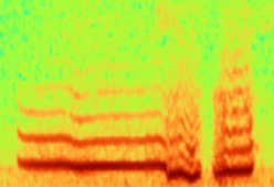
Wheezing
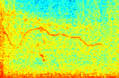
Spectrogram of a wheeze captured by the RESP® Biosensor
Wheezing, a common COPD exacerbation lung sound, is a high-pitched whistling sound indicating partially blocked airways. It is typically heard through exhalation and can be heard with or without a stethoscope or wearable device.
In COPD, airways are obstructed due to inflammation, swelling, mucus or damage in the lungs. Wheezing is a sign of this airway obstruction and occurs when patients are particularly symptomatic due to a flareup, irritant or exacerbation.
Wheezing also occurs in several conditions other than COPD, including asthma, cystic fibrosis, heart failure and interstitial lung diseases.
Crackles
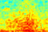
Spectrogram of a crackle captured by the RESP® Biosensor
Crackles, also known as rales, refer to an intermittent rattling, clicking, or popping noise (when small air bubbles move through fluid or mucus) that signifies the presence of mucus or fluid in the airways. They are typically heard during inspiration as the airways re-open and are not alleviated by coughing. These sounds can be heard best in the small and medium airways.
Crackles can be identified when a healthcare professional listens to the sound from a stethoscope or wearable device when the patient breathes in or out.
Crackles can be divided into three separate types:
- Coarse crackles: These crackles are more likely to present in people with COPD. They are lower pitched compared to fine crackles.
- Fine crackles: These are short, high-pitched noises that indicate the presence of fluid in small airways. Doctors often hear this in people with congestive heart failure and pneumonia.
- Biphasic crackles: This sound is a combination of both coarse and fine crackles.
Rhonchi
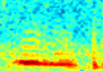
Spectrogram of a crackle captured by the RESP® Biosensor
Rhonchi sounds are low-pitched on the contrary, loud, and coarse, caused by obstruction or narrowing of the airways. Typically heard more during exhalation than inhalation, they often sound like snoring or gurgling and can be confused for wheezing. These sounds are generally heard in the larger airways and can often be cleared with a strong, vigorous cough. Rhonchi can also indicate fluid in the lungs in patients with COPD.
Stridor (A Less Common COPD Lung Sound)
Spectrogram of a stridor
Stridor is a high-pitched, continuous whistle or squeaking sound similar to a wheezing sound due to inflammation in the trachea or larynx. Stridor indicates airway obstruction and can be detrimental if medical attention is not sought. It is typically heard during inhalation but can also be heard during exhalation.
How Are COPD Lung Sounds Assessed?
Auscultation of the lungs with a stethoscope has been the traditional way that clinicians listen to COPD lung sounds. Auscultation takes place during an in-person physical exam and usually lasts for two to three minutes. It typically includes the following steps:
- Patient Positioning: the patient sits or stands comfortably
- Breath Instructions: the patient is asked to take deep breaths
- Listening Points: the clinician places the stethoscope on the patients’ chest and moves downwards. Next, the clinician starts listening on the patients’ back, and moves the stethoscope downward. Symmetrical points should always be compared.
While auscultation has been a valuable approach for diagnosing respiratory disease in COPD patients, it has limitations. Auscultation is episodic, lasting only for a few minutes and requiring patients to visit their doctors in person. It offers clinicians a short window into the patients’ airways which limits the lung sound analysis. Auscultation is also subjective and prone to inter-observer variability.
Innovations in Continuous Auscultation have emerged to address the limitations of episodic auscultation and offer a more complete lung sound analysis. By prescribing a wearable device such as the RESP® Biosensor, clinicians can extend the temporal and geographical range of auscultation and detect abnormal and exacerbation lung sounds in patients’ daily lives. These innovative devices enable clinicians to assess the patient’s response to treatment plans and adjust accordingly for the benefit of the patient’s health — eliminating the need for an in-person visit and potentially reducing a readmission with the appropriate treatment.
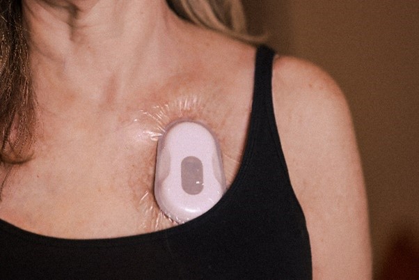
Ask your clinician about the Strados Labs RESP® Biosensor.
Additional Ways to Diagnose and Test for COPD
In addition to identifying abnormal lung sounds, healthcare professionals may also ask patients to perform some of the following tests for identifying COPD:
- Spirometry is a common pulmonary function test (PFT) used to diagnose COPD. Spirometry is used to assess airflow limitations and airflow reversibility. This non-invasive and proven test measures a patient’s forced expiratory volume, indicating lung function and capacity. Spirometry is primarily used to monitor COPD disease progression.
- The Bronchodilator reversibility test combines a pulmonary function test with a bronchodilator. Measuring pre-bronchodilator and post-bronchodilator volumes indicates if the bronchodilator can reverse the symptoms causing the airway restriction or obstruction.
- The Arterial blood gas test measures the carbon dioxide, bicarbonate, pH levels and oxygen levels in the blood. These tests can be conducted in either a hospital or a clinician’s office.
- The Fractional exhaled nitric oxide (FeNO) test measures the amount of nitric oxide in a patient’s breath. High levels of nitric oxide may indicate inflammation of the airways.
- Peak expiratory flow (PEF) is another pulmonary function test that measures how fast a patient can blow out air. This test can be conducted during a spirometry test or with a handheld device.
Frequently Asked Questions
What do normal breath sounds sound like?
Healthy lungs create a smooth, soft sound that is heard when breathing in and out. Normal lung are also called vesicular lung sounds. Hearing normal lung sounds with auscultation means that there isn’t a restriction or obstruction in the airways. The lungs are functioning at full capacity without inflammation or blockage.
What is pleural friction rub? Is it considered a COPD Lung Sound?
Pleural friction rub is an additional lung sound that indicates inflammation of tissues in the pleura area. It is a raspy, grating sound that is less common in COPD and usually occurs in patients with pneumonia, pulmonary embolism, and other diseases.
How is COPD treated and managed?
While there is no cure for COPD, there are several ways to manage and control this chronic disease. These include medications, pulmonary rehabilitation programs, lifestyle changes, oxygen therapy and more.
Is chronic bronchitis a type of COPD?
Yes. Chronic bronchitis is a form of chronic obstructive pulmonary disease involving long-term inflammation of the bronchial tubes. It is characterized by a productive cough lasting more than three months.
What are the most common COPD exacerbation lung sounds?
While every COPD exacerbation is different, the most common COPD exacerbation lung sound is wheezing. Other common symptoms of a COPD exacerbation include productive cough, changes to sputum, dyspnea (shortness of breath) and fatigue.
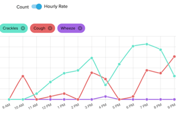
Is coughing common in COPD? What does it mean?
Cough is extremely common in COPD and can have a significant impact on quality of life. With COPD, there is often an overproduction of sputum or phlegm. The body’s coping mechanism is to clear the foreign material from the airway to remove the obstruction. It does this by producing a cough to forcefully remove the secretions. Consistent coughing can also cause airway irritation and inflammation which can result in breathing issues (e.g. labored breathing or shortness of breath). It can also occur during an exacerbation. Cough monitoring technologies have emerged to measure this symptom objectively.
About the Author
Rachel Hollatz, RRT, RPSGT

Rachel Hollatz is the Annotation Manager for Strados Labs, a medical technology company focused on providing clinicians with more objective, reliable data on patients’ respiratory symptoms in the real world. She is a Registered Respiratory Therapist with over 15 years of experience working in health systems including Ministry Health Care and Marshfield Clinic Health System.